Saturday, June 4, 2016

Donut Forest

On the way home from getting donuts this morning Henry asked if we were going to the donut forest...heck yeah we are! Another perfect toddlerism. 


Friday, February 12, 2016

A trick of the light

Sometimes we can be transported to another time and space while performing the most mundane tasks. By turning the polarizer just so I serendipitously stumbled upon these gorgeous landscapes. Glittering and mesmerizing. No longer merely histologic sections of lung or even debris at the edge of a slide. I see aerial views of planets and nighttime cityscapes and topography as seen through an airplane window.

What do you see?


Tuesday, August 18, 2015

Upper esophageal nodule...What is it?

Spotted a small nodule (0.4 cm) in the upper esophagus with pinpoint central erosion...


Below the strip of squamous epithelium, which is focally ulcerated with patchy inflammation, is a circumscribed collection of cardiac-fundic-type glands comprised by parietal (plump pink) and chief cells along with scattered foveolar-type epithelium (not shown here). 


Turned out to be ectopic gastric mucosa (stomach tissue) or ...

Gastric heterotopia of the upper esophagus!


Also called "inlet patch", this lesion is thought to be congenital and is usually located near the upper esophageal sphincter. The majority are asymptomatic and incidentally found; however, gastric acid secretion (those darn parietal cells!) is possible and dysphagia reported by some. Rare complications can include stricture, perforation, and, rarer still, adenocarcinoma.

The decedent died of hypertensive and atherosclerotic cardiovascular disease with evidence of remote infarct with potential acute component (histology pending).  Cardiac work-up a few months prior to death was inconclusive and the patient continued to complain of and self-treat "heartburn" symptoms.  Possible that heart was entirely the source of the symptoms misinterpreted as heartburn.  Also possible that, although quite small, the lesion could have contributed to the symptoms the decedent experienced in months prior to his death. 


Thursday, May 7, 2015

Art from Artifact

Beauty in the unexpected

In pathology, an artifact is an artificial change introduced as a result of some step in tissue manipulation. Tissue processing -progressive dehydration and paraffinization - produces reproducible "artifacts" as a result of inherent properties of some tissues lending their "classic" appearance under the microscope (e.g. "fried egg" appearance of oligodendrocytes, "orphan Annie-eye" nuclei in follicular thyroid carcinoma).  In general, though, artifact is something to be avoided -an unwanted result of some misstep in the tissue handing process...improper fixation, air drying, or the dreaded air bubble under the coverslip.  These artifacts can physically obscure the tissue and even alter the appearance of cells.  Sometimes the effect is small like tiny flecks of black formalin pigment in the background.  At its worst, artifact can make tissue sections nearly uninterpretable (e.g. cautery artifact or "cooked" tissue, ice crystals in frozen section).

As I was looking at sections of frontal cortex today for a research study, many of the slides had fallen victim to the plight of too little mounting medium causing air bubbles everywhere!! These bubbles produce black masses (highly refractile without the condenser) under the microscope obscuring the tissue beneath. As I was fighting with the bubbles, I began to notice the amazing arrangements they made as the air dissected along the tissue surface -these beautiful ice crystal-like organic structures forming tiny forests on the microscopic surface of neurons, glia, dendrites, axons, capillaries and arterioles...little rivers formed by negative spaces between gyri.

More strange little planets and unexpected landscapes.

Here's to finding the unexpected beauty in the artifacts of life.


Monday, February 9, 2015

Wriggly worm

Henry met his first earthworm today. The poor guy was wriggling on the doormat as we walked up to ring the doorbell at daycare. (sidenote: this is a common hazard of rainy Seattle weather as you often have to watch your step for beached worms). "We" placed him in the grass, rescuing him from almost certain demise. As we did, Henry tried out his new word, "wohm!".  When Faduma opened the door, he exclaimed "Wohm, Wohm!" and then said "Thank you, Mama!" ...and my heart melted.

That is all. Have a good night :)


Monday, February 2, 2015

Magpie in the house

Henry has turned into a regular magpie, pilfering and stashing treasures in all kinds of hidden places. Things mysteriously go missing and then I remember, "oh yeah, we have a toddler in the house".  Then it all makes sense in the way that toddlers make no sense at all, being the completely unpredictable creatures that they are. Latest missing item...my hairbrush. Eh well, that's what pony tails are for. Here's to life with silly toddlers and the magic they bring. Thanks for making our lives so interesting.

I love you my little magpie.


Saturday, December 13, 2014

Up in the sky!


A little fun with toddler plane-spotting. Original photo courtesy of Dad.

Our son is obsessed with pointing out each plane that flies overhead. "MaHn!!", he exclaims. I'll always cherish those sweet first words.

Sunday, December 7, 2014

Seas of pink


Diffuse, neuritic, dense core, fibrillary, primitive, compact, burned-out, cotton wool, fleecy, lakes -all ways to describe the morphologies of amyloid beta, the plaque-forming pathologic protein of Alzheimer's disease, in the brain. 


200X HE/LFB. Case with severe Alzheimer's pathology with back-to-back amyloid plaques comprising the majority of the field.  These dense, pink, fluffy spherical aggregates are surrounded by islands of vacuolated brain tissue with significant reactive astrocytosis (cells with plump, bright pink cytoplasm and visible processes).  


Reminds me of fluffy cotton candy...


Friday, December 5, 2014

Build a brain

Before we start, a little background...

At least 6 months of my second (and final) year of neuropathology fellowship focuses on the neuropathological work-up of dementia. The most common types we encounter are Alzheimer's disease, vascular dementia, and Lewy Body dementia; however, we have our fair share of other diverse entities including rare genetic disorders.
  

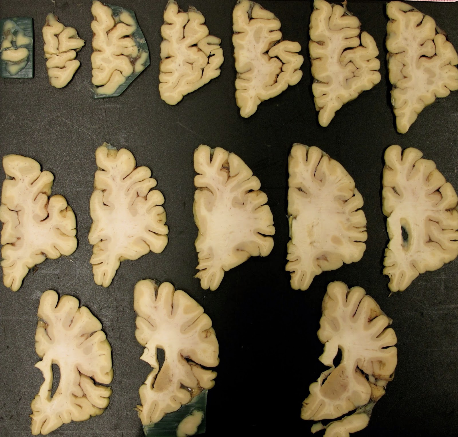
Our process involves soaking the brain in formalin fixative for at least 10 days to preserve the tissue. The brain is then embedded in a gelatin mold to stabilize the tissue for cutting into thin (3 to 4 mm thick), uniform sections. These sections are then laid out in order from front (anterior) to back (posterior) across several boards. 


The process of evaluating all aspects of the tissue that can be seen with the naked eye, followed by sampling of representative standard sections (those required to diagnose the three most common types of dementia noted above as well as other pathology) as well as any abnormal areas, is called gross examination or "grossing" the tissue.

4 mm thick coronal brain slices (anterior ~1/3 of right side of brain)


The sampled tissues are then processed, put onto glass slides, and stained for examination under the microscope. Sampling only accounts for probably ~5% of the brain tissue. Remaining tissues are stored in tidy glass jars as a part of our repository.


What happens if you have to go back to the previously cut and sampled brain? It is not infrequent in pathology that a specimen needs to be revisited for one reason or another in order to submit additional tissue. That is when the fun starts!


Previously sampled brain slices enjoy great freedom and entropy upon release from their jar.


For this particular case, I needed to re-examine possible lesions on the surface of the posterior, inferior frontal lobe and the posterior, superior temporal lobe. The best way to fully evaluate the surface again is to reconstruct the left half of the brain. Even though I only needed to put the anterior half together, I thought...why stop there? 


Reconstructed left half of brain (all cortical slices accounted for except for 3 overly fragmented slices)


I am in constant awe at the beauty of the human brain. Each case is a bit different and reveling in these structures never gets old.


I highly recommend reconstructing a brain to really hone your neuroanatomy skills! 


Thursday, December 4, 2014

Cerebellum V1

Put a bird on it...


           Dark-eyed Junco atop cerebellar Purkinje cell dendritic arbor (Calbindin IHC, 200X)


Saturday, November 15, 2014

Tiny Worlds

The stark black border of photos taken through the microscope with the crisp outline of the ocular and it's vivid contents imparts a sense of depth and utter "largeness" conjuring visions of far away and undiscovered planets. 


Seeing Spots


Bielschowsky silver stain. 400X magnification.
Aggregates of corpora amylacea in leptomeninges and molecular layer of cortex


Alpha-synculein IHC (for Lewy body [LB] detection -no LB's present). 400X magnification.
Numerous corpora amylacea in superficial cortex


Sidenote: Corpora amylacea (coined by Virchow in 1854 for appearance resembling starch granules) are polyglucosan  (carbohydrate-based) bodies that accumulate in the CNS with age and can be found within astrocytic processes and axons. These were found in the brain of a 90 year-old without cognitive impairment.

Sunday, April 10, 2011

Remembrance of Things Past

This is the site of my most cherished childhood memories.  A little green and gold house on Settles street in Big Spring, Texas. This unassuming home belonged to my great Aunt and Uncle.  This couple, unable to have children of their own together, gave so much to the children of others.  My siblings and I basically grew up here, wanting to spend every moment we could in this home so full of fun, laughter, whimsy, imagination, love and safety. Everyone that knows me is aware that the childhood of myself and my siblings was not easy, but time spent in this house, with two incredible people, made that all dissolve into the background.  

Aunt Pat and Uncle Scotty passed away in 2006.  This post is about trying to cling to memories that made me who I am today.


Belongings from our loved ones act as tangible reminders of their presence on earth, and it is human nature to want to hang on to these things.  Sadly, in order to obtain these precious talismans, my Aunt and I found ourselves at the estate sale of the green and gold house on Settles Street.  


I wish that I could accurately describe the feeling of going through your childhood sanctuary with strangers, pilfering through that which you hold so dear.  Realizing that many of the things you had your heart set on keeping had already be taken.  Unnatural is most fitting. Surreal. 


These are the treasures I rescued.


I realized when I got them all home that, out of that house, without my Aunt and Uncle, these objects had lost some of their magic.  You can never really go back home as they say. Still, though they are merely trinkets, they are my treasures and are more special to me than I can ever truly say.


This is dedicated to Aunt Pat and Uncle Scotty. Thank you for the laughs, and most importantly, all the love.  You are sorely missed.


*Thanks to Marcel Proust for the title...I will finish In Search of Lost Time someday