Before we start, a little background...
At least 6 months of my second (and final) year of neuropathology fellowship focuses on the neuropathological work-up of dementia. The most common types we encounter are Alzheimer's disease, vascular dementia, and Lewy Body dementia; however, we have our fair share of other diverse entities including rare genetic disorders.
Our process involves soaking the brain in formalin fixative for at least 10 days to preserve the tissue. The brain is then embedded in a gelatin mold to stabilize the tissue for cutting into thin (3 to 4 mm thick), uniform sections. These sections are then laid out in order from front (anterior) to back (posterior) across several boards.
The process of evaluating all aspects of the tissue that can be seen with the naked eye, followed by sampling of representative standard sections (those required to diagnose the three most common types of dementia noted above as well as other pathology) as well as any abnormal areas, is called gross examination or "grossing" the tissue.
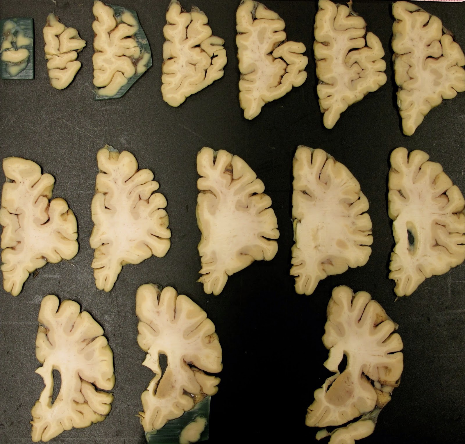 |
4 mm thick coronal brain slices (anterior ~1/3 of right side of brain)
The sampled tissues are then processed, put onto glass slides, and stained for examination under the microscope. Sampling only accounts for probably ~5% of the brain tissue. Remaining tissues are stored in tidy glass jars as a part of our repository.
What happens if you have to go back to the previously cut and sampled brain? It is not infrequent in pathology that a specimen needs to be revisited for one reason or another in order to submit additional tissue. That is when the fun starts!
|
 |
Previously sampled brain slices enjoy great freedom and entropy upon release from their jar.
|
For this particular case, I needed to re-examine possible lesions on the surface of the posterior, inferior frontal lobe and the posterior, superior temporal lobe. The best way to fully evaluate the surface again is to reconstruct the left half of the brain. Even though I only needed to put the anterior half together, I thought...why stop there?
 |
Reconstructed left half of brain (all cortical slices accounted for except for 3 overly fragmented slices)
|
I am in constant awe at the beauty of the human brain. Each case is a bit different and reveling in these structures never gets old.
I highly recommend reconstructing a brain to really hone your neuroanatomy skills!


